The 19 Muscles Of The Foot : Practical (Leg Model) at Tidewater Community College ... / They are usually but little developed, and, being situated in masses, it is difficult to isolate and describe distinctly the precise action of 19.
byAdmin•
0
The 19 Muscles Of The Foot : Practical (Leg Model) at Tidewater Community College ... / They are usually but little developed, and, being situated in masses, it is difficult to isolate and describe distinctly the precise action of 19.. Don't forget to utilise these top anatomy study tips! They are generally divided into two sets: Contrary to expectations, the intrinsic foot muscles contribute minimally to supporting the arch of the foot during walking and running. The muscles are located mainly in the sole of the foot and divided into a central (medial) group and a group on either side (lateral). Flexion of 4 lesser toes at metatarsophalangeal, proximal & distal interphalangeal joints inversion of foot plantar flexion of ankle.
Interossei refer to muscles between certain bones. The foot owes its peculiarities of form to its bony structure; We derive a dimensionless curvature parameter that governs the stiffness contribution of the transverse tarsal arch, demonstrate its predictive power using mechanical. Models of foot function explore different models of foot function with podiatrist kevin bruce powered by physiopedia start course presented by: Those of the medial plantar region are connected with the great toe, and corrrespond with those of the thumb;
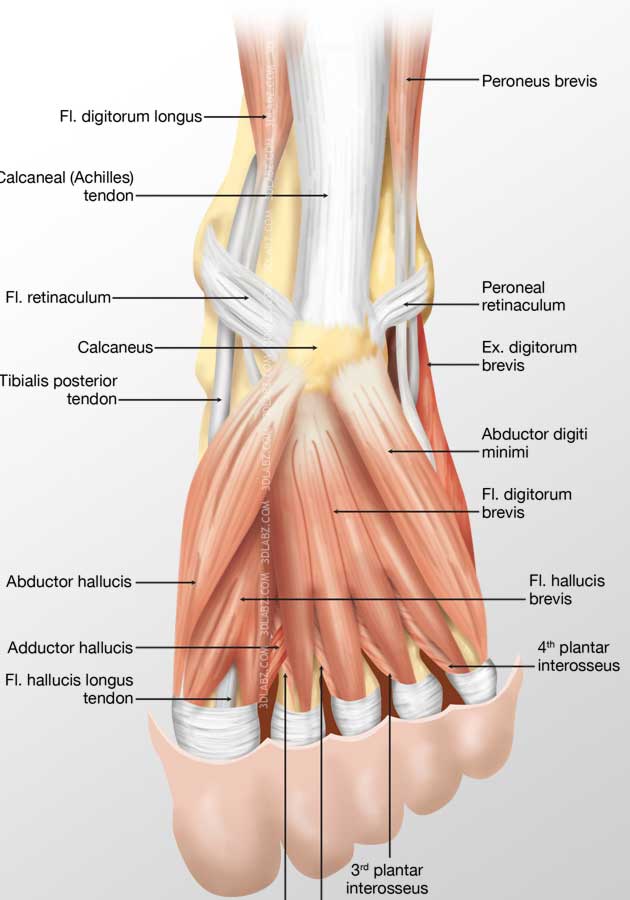
Foot Dorsal Muscles 3D Illustration | Price from www.3dlabz.com The interosseous muscles of the foot are muscles found near the metatarsal bones that help to control the toes. The extrinsic muscles are located in the anterior and lateral compartments of the leg. Medial and lateral processes of posterior calcaneal tuberosity. Their limited impact on posture and movement has led to the broad use of the extensor hallucis brevis and extensor digitorum brevis as muscular sources for tissue grafts. Deep intrinsic muscles of the foot through video. Calluses are created by friction applied to the skin of the foot, often by misfitting shoes. Muscles & tendon sheaths of the foot. Don't forget to utilise these top anatomy study tips!
The muscles round off the angular structure.
(10 foot/ankle and 19 intrinsic) ten of these muscles originate outside of the foot itself but the other 19 muscles are referred to as intrinsic muscles of the foot and act only within the foot. They include the abductor halluces, the flexor digitorum brevis, the abductor digiti minimi, and the quadratus plantae. This is an online quiz called muscles of the foot. The dorsal aponeurosis of the toes supports the effect of the dorsal foot muscles by redirecting the force line of their tendons to. Muscles of the foot laminated anatomy chart. Your feet work tirelessly day in and day out. The muscles are located mainly in the sole of the foot and divided into a central (medial) group and a group on either side (lateral). Related online courses on physioplus. Your foot and ankle specialist can shave down the thick layers of a callus using a scalpel blade. Most are located on the inferior part of the foot. This article outlines the basic anatomy of the foot bones. All intrinsic muscles of the foot are innervated by branches of the tibial nerve except for extensor digitorum brevis, which is innervated by the deep. Deep intrinsic muscles of the foot through video.
The four lumbricales are affixed to the inner side of the four toes. Interossei refer to muscles between certain bones. This article outlines the basic anatomy of the foot bones. When the muscles tighten (contract). All intrinsic muscles of the foot are innervated by branches of the tibial nerve except for extensor digitorum brevis, which is innervated by the deep.
Muscles of Leg and Foot - Anatomy 3300 with Derek Harmon ... from s3.amazonaws.com They are usually but little developed, and, being situated in masses, it is difficult to isolate and describe distinctly the precise action of 19. There is a printable worksheet available for download here so you can take the quiz with pen and paper. Layer 3 of the foot. Your foot and ankle specialist can shave down the thick layers of a callus using a scalpel blade. The muscles round off the angular structure. The muscles are located mainly in the sole of the foot and divided into a central (medial) group and a group on either side (lateral). Flexor hallucis longus tendon transfer to the dorsum of the foot and release of the flexor digitorum longus and brevis tendons at the base of each toe. Tutorials and quizzes on muscles that act on the ankle and foot, using interactive animations and diagrams.
Learn and reinforce your understanding of sole:
Tutorials and quizzes on muscles that act on the ankle and foot, using interactive animations and diagrams. The muscles acting on the foot can be divided into two distinct groups; Muscles of the ankle and foot. They are generally divided into two sets: Base of the 5th metatarsal. The interosseous muscles of the foot are muscles found near the metatarsal bones that help to control the toes. Most are located on the inferior part of the foot. Those of the medial plantar region are connected with the great toe, and corrrespond with those of the thumb; Models of foot function online course: An overview of the intrinsic muscles of the foot including their origin, insertion, blood supply, innervation, function and clinical relevance. The tendons are thick bands that connect muscles to bones. They include the abductor halluces, the flexor digitorum brevis, the abductor digiti minimi, and the quadratus plantae. This article outlines the basic anatomy of the foot bones.
Interossei refer to muscles between certain bones. Flexor hallucis longus tendon transfer to the dorsum of the foot and release of the flexor digitorum longus and brevis tendons at the base of each toe. Layer 3 of the foot. Calluses are created by friction applied to the skin of the foot, often by misfitting shoes. Contrary to expectations, the intrinsic foot muscles contribute minimally to supporting the arch of the foot during walking and running.
foot anatomy | Muscular and Skeletal Anatomy of Ankle and ... from i.pinimg.com The foot owes its peculiarities of form to its bony structure; Anatomy warehouse is the largest supplier of anatomy models and healthcare education models to the musculature of the head anatomy chart is a very comprehensive and beautifully illustrated view of the muscles that make up our head, parts of. The muscles acting on the foot can be divided into two distinct groups; There are over two dozen. We derive a dimensionless curvature parameter that governs the stiffness contribution of the transverse tarsal arch, demonstrate its predictive power using mechanical. When the muscles tighten (contract). Models of foot function explore different models of foot function with podiatrist kevin bruce powered by physiopedia start course presented by: Learn and reinforce your understanding of sole:
The foot owes its peculiarities of form to its bony structure;
The muscles covered in this article serve various. The skeleton of the foot is often subdivided, based on functional and clinical 10.16 the short muscles of the right foot from the plantar view. Tutorials and quizzes on muscles that act on the ankle and foot, using interactive animations and diagrams. The four lumbricales are affixed to the inner side of the four toes. The interosseous muscles of the foot are muscles found near the metatarsal bones that help to control the toes. Contrary to expectations, the intrinsic foot muscles contribute minimally to supporting the arch of the foot during walking and running. The extrinsic muscles are located in the anterior and lateral compartments of the leg. There are 29 muscles associated with the human foot. Medial and lateral processes of posterior calcaneal tuberosity. Calluses are created by friction applied to the skin of the foot, often by misfitting shoes. The foot owes its peculiarities of form to its bony structure; However, these muscles do influence our ability to produce forward propulsion from one stride into the next, highlighting their role in bipedal locomotion. Do you know what major muscles extend the foot?